
Wood anatomy
More than three trillion trees live on earth (Crowther et al. 2015). During the live and growth they continuously record environmental information in their cell structure, tree-ring width, wood density and chemical composition. Due to their long live spans from decades to millennia, trees are ‘environmental information systems’ in space and time. The information from tree-rings help to estimate carbon and evapotranspiration dynamics of forest ecosystems (Babst et al. 2014, Frank et al. 2015), mortality waves (Park Williams et al. 2013), climate variations in the past millennia (Masson-Delmotte et al. 2013) or the reaction of tree species to the expected challenges of global change (Charney et al. 2016). Until now, the most common parameter used is classical tree-ring width, which is a single integrated measure of radial tree growth over one growing season.
However, tree-ring width is not the ideal measure for estimates of tree’s and tree species’ reactions to the now more frequent extreme events like droughts or late frosts (Diffenbaugh et al. 2017), insect calamities or to study fundamental seasonal dynamics in a world with a changing climate. To study such topics, better measure with a higher temporal resolution and a closer link to the physiological mechanisms within trees are needed. Wood anatomical studies within tree-rings, i.e. high temporal resolution measures of the continuous wood formation, are a promising new field. On the one side, the higher temporal resolution allows a better correlation of stressor and stress signal (e.g. short time flooding and the following decline of tree growth). On the other side, wood anatomical parameters like cell (tracheid/vessel) diameter are directly linked to physiological processes like water transport (for example drought stress is related to cell diameter), which is an advantage over classical tree-ring width.
As a result, wood anatomical parameters are often more strongly correlated to climate variables than tree-ring width. Furthermore, the different wood anatomical parameters within a tree-ring (lumen area, cell-wall thickness, number of cells per area) provide additional information because they oft respond to different climatic or environmental signals then tree-ring width (Fonti et al. 2010).
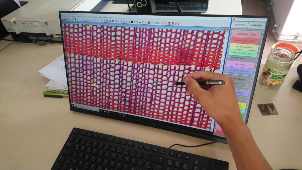
The basic methods for wood anatomical studies are the preparation of thin sections from tree cores or the continuous collection of short (micro) cores for the monitoring of tree growth. The thin sections from the cores are than photographed with a microscope (classical or confocal laser microscopy) and digitized. These digital images then have to be analyzed with software like WinCell, ROXAS or ImageJ, which all work semi-automatically with custom settings for each species (von Arx et al. 2016, von Arx and Carrer 2014).
The theoretical foundations for wood anatomical studies are thus established, but the current methods are very time and labor intensive. Currently the established methods are only well developed for a few tree species, mostly conifers and a fully automatic analysis of the images is not possible yet. Because of this there is a lack of long chronologies of wood anatomical parameters, chronologies of less common species or chronologies form the various possible site conditions. Wood anatomical studies of forests and trees with a large spatial and temporal extent have thus not been possible yet.

This is where DIG-IT! takes action. A fully automatic assessment of wood anatomical parameters like cell diameter, cell-wall thickness or mark rays of the most important tree species in Mecklenburg-Vorpommern appears possible by applying DCNNs (Garcia-Pedrero 2018). This promises new insights regarding the resistance and resilience of native and non-natiave tree species towards extreme climatic events, the stability of forest ecosystems with respect to forest management options, and the climatic variability of the past centuries to millennia. The world wide growing field of quantitative wood anatomy would greatly benefit from a time-saving automatic analysis of wood anatomical images and experience a large development boost.
The project "DIG-IT!" is funded by the European Social Fund and the Ministry for Education, Science and Culture of the federal state Mecklenburg-Vorpommern.